
My dog has a torn CrCL – what do I need to know?
Rupture of the cranial cruciate ligament (ACL) is a result of too much stress/force being applied to a genetically weak ligament. Damage to the ligament itself, and the knee cartilage (meniscus) occurs allowing the knee to “slide” rather than “hinge.” We call this sliding motion cranial “drawer sign.” (Think of pulling out a dresser drawer.)
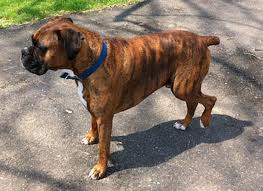
This can occur in any breed but breeds that seem to be predisposed are Boxers, German Shepherds, Labradors, Golden Retrievers, Newfoundlands, Mastiffs, and Akitas. Young and old dogs can be affected but most commonly affected are middle aged to older dogs. Can present as an acute or chronic lameness that will not improve with rest and NSAID (non-steroidal anti-inflammatory drug) therapy.
Orthopedic exam findings:
(sedation may be necessary to complete an accurate orthopedic exam)
- May sit with knee rotated outward, called a positive sit test
- Positive for cranial drawer sign (may not appreciate if a partial tear present or if animal is fighting palpation)
- Positive tibial thrust
- Painful on extension
- Presence of medial buttress
- this is an accumulation of fibrous scar tissue that the body deposits in an effort to stabilize the knee
- this is an indicator of how chronic this condition is, along with muscle atrophy.
- Palpable joint effusion (swelling)
- Possible meniscal “click” if the meniscus is damaged
Radiograph findings:
- Osteophytes (bone spurs) typically present along femoral trochlear ridge, tibial plateau, and patella
- Effusion (accumulation of inflammatory mediators) seen in joint space
Treatment options:
Surgical repair is aimed at stabilizing the knee to prevent the sliding motion that causes lameness. Recovery time from surgery is 8-16 weeks depending on the surgery performed.
- Lateral suture – “Extra-cap” (extracapsular repair using monofilament)
- Heavy suture (usually 40, 80, or 100lbs test monofilament) placed around the fabella on the lateral aspect of femur and through a hole in the tibia. This runs in similar orientation to the original cranial cruciate ligament. Except, your prosthesis is outside the joint. “Extracapsular repair.”
- Usually done on smaller dogs and less active breeds
- Recovery time 6-8 weeks
- Tibial Plateau Leveling Osteotomy – “TPLO”
- Osteotomy (cutting into bone) is performed at the proximal tibia and the tibial plateau is then rotated and stabilized with a bone plate. This biomechanically changes the leg such that there is more load on the caudal cruciate ligament (which can handle the load) and eliminates tibial thrust.
- The cranial cruciate ligament is not fixed. You will always have cranial drawer sign. However, the leg will be dynamically stable.
- Recovery time 12-16 weeks
- Tibial Tuberosity Transposition – “TTA”
- Cut along cranial aspect of proximal tibia
- Insert cage in between the tibia to advance it
- Goal is to make the patellar tendon perpendicular to the tibial plateau.
- This results in elimination of tibial thrust, similar to TPLO
- Newer procedure, early results comparable to TPLO thus far
- Recovery time 12-16 weeks
Surgery remarks:
- Most surgeons consider the TPLO as the gold standard for cruciate repair, although this is controversial. The extra-cap was considered the gold standard for a long time. Results of patients a year after surgery are comparable between the TPLO and extra-cap.
- Any surgical repair can still result in progression of osteoarthritis. However, the surgically repaired joint will have a much slower progression than a non-surgically repaired joint. The patient’s return to normal activity is much sooner with surgery compared to medicine and cage rest.
Cage Rest with use of NSAIDs:
- This may work with small dogs, but they need strict confinement
- Typically 8-12 weeks
- If surgery is unaffordable, this is the next best option. The patient will eventually develop fibrosis that will help stabilize the leg. The progression of osteoarthritis will be greater and occur sooner, compared to surgical repair.
Facts to Know:
- Very high chance they will rupture the cruciate ligament on the other leg in the next 12 months (following the initial ligament injury). Studies say there is between a 60-90% chance.
If you think your dog may have torn their CrCL, please call us for an appointment.